Journal of Spine Research
Online ISSN : 2435-1563
Print ISSN : 1884-7137
Current issue
Displaying 1-19 of 19 articles from this issue
Editorial
Review Article
  • Koichiro Ide, Yu Yamato, Tomohiko Hasegawa, Go Yoshida, Tomohiro Banno ...
    2025 Volume 16 Issue 6 Pages 819-825
    Published: June 20, 2025
    Released on J-STAGE: June 20, 2025
    JOURNAL FREE ACCESS

    The TOEI study is a cohort study of physical examinations that have been conducted every two years since 2012 as part of the community health examinations in Toei Town, Aichi Prefecture. The most significant feature of this study is that it is possible to accumulate longitudinal data over 10 years. Whereas most studies have been limited to cross-sectional studies, the TOEI study is taking advantage of the strengths of longitudinal research, allowing the analysis of changes over time.

    This report emphasizes that changes of spinal alignment in elderly people were observed earlier in women than in men. In addition, the progression of low back pain was found to be related to spinopelvic parameters and motor function decline. Kyphosis of spinal alignment was more common in the upper lumbar spine, and the sacral slope and inflection point were found to be risk factors over time.

    Through these new findings based on long-term data, we will continue to contribute to the prevention and treatment of low back pain and of spinopelvic alignment deterioration.

    Download PDF (1213K)
  • Masashi Uehara, Jun Takahashi, Shota Ikegami, Ryosuke Tokida, Hikaru N ...
    2025 Volume 16 Issue 6 Pages 826-830
    Published: June 20, 2025
    Released on J-STAGE: June 20, 2025
    JOURNAL FREE ACCESS

    Introduction: We established the Obuse Study, involving a cohort of local residents, to clarify the degree and frequency of age-related changes in the musculoskeletal system. The objective of this study was to investigate the sagittal alignment of the spinal column to clarify: 1) the characteristics of spinal column alignment by sex and age group; 2) the relationship between sagittal alignment and motor function; and 3) the frequency of low back pain and factors related to low back pain.

    Methods: Invitations for examinations were sent to 1,297 randomly selected residents of Obuse, Nagano Prefecture, who were randomly selected from 5,352 residents aged 50-89 years listed in the resident registry. A total of 413 people (approximately 50 men and 50 women per age group) underwent examinations of motor function and standing full-spine lateral radiographs. Spinal sagittal parameters were measured. Their characteristics were analyzed by age and sex, and the relationship between spinal sagittal alignment and motor function (knee extension-flexion muscle strength, time to stand on one leg, grip strength, and locomotion test) and their relationships with low back pain were examined.

    Results: Changes in spinal sagittal alignment were more pronounced in men, with an anterior shift of the cervical spine occurring at age 60 years and older. In women, decreased lumbar lordosis and pelvic retroversion occurred at younger ages than in men. The worse the sagittal alignment of the spine, the lower the performance in grip strength, stand-up, and two-step tests. Low back pain was present in 12.9% of the participants, and pelvic incidence-lumbar lordosis (PI-LL) mismatch (>10 degrees) was an independent and significant factor associated with low back pain.

    Conclusions: A cross-sectional study of sagittal spinal alignment in a community-dwelling population showed that changes in sagittal spinal alignment tended to differ between men and women. There was a clear association between sagittal spinal alignment and physical function. The approach to sagittal spinal alignment may be an important disease control measure not only for low back pain in elderly persons, but also for motor function decline.

    Download PDF (841K)
  • Tadatsugu Morimoto, Hirohito Hirata, Masatsugu Tsukamoto
    2025 Volume 16 Issue 6 Pages 831-836
    Published: June 20, 2025
    Released on J-STAGE: June 20, 2025
    JOURNAL FREE ACCESS

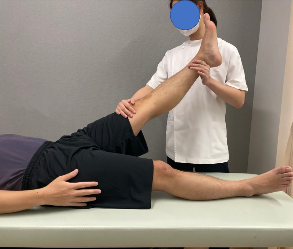
    Low back pain is common and has a wide range of causes, including spinal, pelvic and hip lesions and visceral lesions. Although infrequent, some conditions are associated with abdominal aortic disease or paralysis requiring urgent response, hence an accurate differential diagnosis is essential in the outpatient treatment of low back pain. Thus, it is important to recall the appropriate differential diagnosis from a careful medical interview and to gather evidence from physical examination findings to support the diagnosis. This article describes the interview and physical findings in outpatient low back pain care, with particular attention to the SLR test.

    Download PDF (1112K)
  • Michio Hongo, Akira Saito, Yuji Kasukawa, Naohisa Miyakoshi
    2025 Volume 16 Issue 6 Pages 837-842
    Published: June 20, 2025
    Released on J-STAGE: June 20, 2025
    JOURNAL FREE ACCESS

    Exercise therapy plays a crucial role in functional recovery and improving patients' quality of life in the treatment of low back pain. Orthopedic surgeons diagnose the cause of low back pain through examinations and prescribe appropriate exercise therapy, but collaboration with physical therapists is essential for effective implementation. Orthopedic surgeons identify the cause of low back pain through patient interviews, physical examinations, and imaging studies to determine the most appropriate treatment. In exercise therapy, personalized prescriptions based on an accurate diagnosis are required. However, evidence supporting exercise therapy tailored to specific causes of back pain remains limited, posing a challenge for future research. Meanwhile, the effectiveness of exercise therapy for chronic low back pain is recommended in clinical guidelines, with aerobic exercise and core stabilization training being recognized as beneficial. Physical therapists, based on the diagnosis provided by orthopedic surgeons, conduct detailed evaluations and implement tailored treatments for patients. They utilize a combination of manual therapy, stretching, and core muscle training to alleviate back pain. Additionally, they contribute to the prevention of low back pain through lifestyle guidance and workplace education. Particularly, combining exercise with educational interventions is expected to yield greater preventive effects. However, collaboration between orthopedic surgeons and physical therapists faces challenges such as insufficient information sharing and issues with treatment continuity. To address these problems, utilizing ICT for real-time communication, organizing conferences, and implementing interdisciplinary training programs can be effective strategies. Strengthening cooperation at every stage of diagnosis, treatment, and education is essential for enhancing the effectiveness of low back pain management.

    Download PDF (1195K)
Original Article
  • Tomoyuki Osato, Noriaki Kawakami, Toshiki Saito, Nobuchika Kawasaki, N ...
    2025 Volume 16 Issue 6 Pages 843-848
    Published: June 20, 2025
    Released on J-STAGE: June 20, 2025
    JOURNAL FREE ACCESS

    With the increasing elderly population, the number of patients with spinal deformities accompanied by low back pain is rising, highlighting the importance of conservative treatments alongside surgical options. So, we've providing short-term hospitalization with torso cast fixation and physical therapy, followed by torso brace treatment after discharge, for adult spinal deformities. This study included 19 patients (mean age 75.3 years; 4 males, 15 females) with adult spinal deformities or adult degenerative scoliosis, all showing spinal flexibility in the supine position. Torso cast fixation was performed using a Risser table, and dynamic gait analysis was conducted before and after the intervention using the VICON system. Single support slightly decreased, while cadence, step length, stride length, walking speed, and hip joint angle showed improvement trends, though without statistical significance. Gait analysis videos revealed notable improvements after torso cast application and brace use. These findings suggest that torso casts and braces provide effective spinal support, contributing to both static and dynamic gait improvements.

    Download PDF (1390K)
  • Megumu Kawai, Satoshi Kato, Noriaki Yokogawa, Yuki Tanaka, Takaki Shim ...
    2025 Volume 16 Issue 6 Pages 849-854
    Published: June 20, 2025
    Released on J-STAGE: June 20, 2025
    JOURNAL FREE ACCESS

    Introduction: In Japan, now a super-aging society, the proportion of elderly patients undergoing spinal surgery continues to increase. Reduced bone density has been reported to be a risk factor for postoperative complications such as screw loosening and intervertebral cage subsidence. Recently, preoperative osteoporosis screening and treatment have been emphasized to prevent postoperative complications in spinal fusion surgeries. However, the intervention and screening are often insufficient. This study investigated the prevalence of osteoporosis and the current treatment status in patients with lumbar spinal stenosis (LSS) undergoing surgeries. It also examined the problems of osteoporosis treatment in patients with LSS.

    Methods: A total of 145 patients who underwent decompression of up to two levels or single-level fusion for LSS at our institution during the period from January 2016 to December 2023 were included. Preoperative bone density was measured using dual-energy X-ray absorptiometry (DEXA) of the lumbar spine and proximal femur. Existing vertebral fractures were examined by whole-spine X-ray. Osteoporosis was diagnosed according to the Japanese guidelines. The prevalence of osteoporosis was examined. Preoperative and postoperative treatment statuses were compared between male and female patients.

    Results: Overall, 21.4% of the patients were diagnosed with osteoporosis (14.6% in men, 28.5% in women). Only 2.1% of patients had a lumbar DEXA value ≤70%. Only a few patients were diagnosed with osteoporosis by lumbar DEXA. The rate of osteoporosis treatment intervention remained low in men both before and after surgery.

    Conclusions: Patients with LSS had a higher prevalence of osteoporosis than the general population. In particular, male patients had insufficient treatment, indicating a need for more proactive management of osteoporosis in this population. In addition, the diagnosis of osteoporosis was difficult using only lumbar DEXA in this population due to degeneration.

    Download PDF (981K)
  • Yoshinobu Hagihara, Fumio Hasue, Atsushi Terakado
    2025 Volume 16 Issue 6 Pages 855-859
    Published: June 20, 2025
    Released on J-STAGE: June 20, 2025
    JOURNAL FREE ACCESS

    Introduction: Patients with low back pain underwent caudal block (CB) and were classified into two groups based on their Pain Detect (PD) scores, with the aim of clarifying the effectiveness of CB.

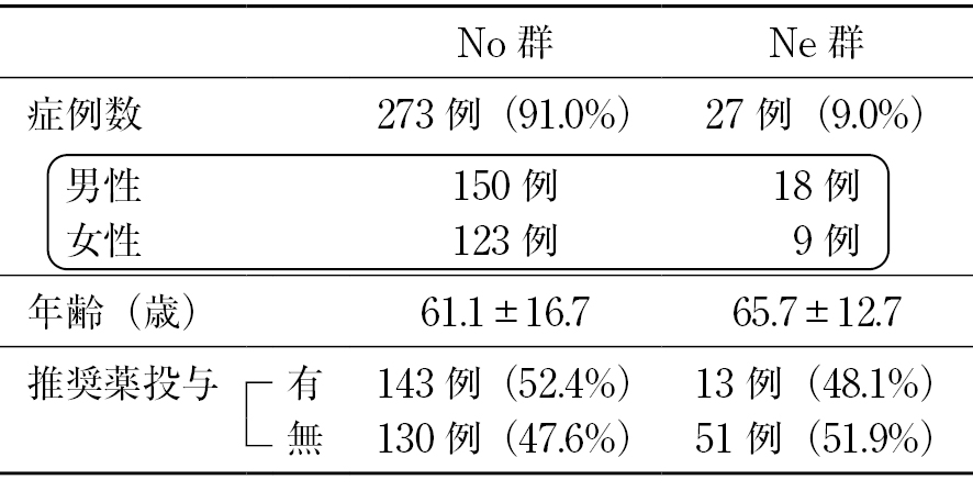
    Methods: From May 2019 to July 2024, 532 sessions were conducted for 300 patients with low back pain who underwent CB and had their properties evaluated using PD. They were then grouped by their PD scores: non-neuropathic pain group (No group), with PD score < 19 points; and neuropathic pain group (Ne group), with PD score ≥19 points. The items examined were the visual analog scale (VAS) scores before and 1 hour (1 h), 1 day (1 d), and 1 week (1 w) after CB, and the VAS improvement rate at 1 h, 1 d, and 1 w after CB.

    Results: There were 273 cases (91.0%) in the No group and 27 cases (9.0%) in the Ne group. The VAS was significantly lower in the No group than in the Ne group throughout the entire course, and the VAS improvement rate was significantly greater in the No group than in the Ne group throughout the entire course. Within the groups, the VAS at 1 h, 1 d, and 1 w was significantly lower than before.

    Conclusions: In treating low back pain, though CB is more effective for non-neuropathic pain than for neuropathic pain, it is considered worthwhile to perform it as a treatment.

    Download PDF (854K)
  • Kotaro Aburakawa, Kanichiro Wada, Gentaro Kumagai, On Takeda, Yasuyuki ...
    2025 Volume 16 Issue 6 Pages 860-866
    Published: June 20, 2025
    Released on J-STAGE: June 20, 2025
    JOURNAL FREE ACCESS

    Introduction: Whether recapping laminoplasty can prevention postoperative spinal kyphotic deformity following surgery for spinal tumors is unknown. The purpose of this study was to compare the postoperative spinal kyphotic deformity of lumbar intradural tumors patients who underwent recapping laminoplasty and those who underwent laminectomy.

    Methods: A retrospective study involving 20 patients (9 males, 11 females, mean age: 55.3 years) with lumbar intradural tumors who underwent surgery at our hospital and were treated for more than 2 years after surgery was performed. The patients were divided into two groups: 7 patients who underwent laminectomy (LN group) and 13 patients who underwent recapping laminoplasty (LP group). The progression of postoperative spinal kyphotic deformity from before surgery to 6 months, 1 year and 2 years after surgery was compared.

    Results: Kyphotic deformity at 1 year and 2 years after surgery was significantly smaller in the LP group (1 year: LP group 0.6±2.0, LN group 7.9±5.5, p = 0.008; 2 years: LP group 1.3±2.9, LN group 7.6±4.8, p=0.037). There was no significant progression of kyphotic deformity at 6 months, 1 year, or 2 years after surgery in the LP group.

    Conclusion: Recapping laminoplasty for patients with lumbar intradural tumor is a useful method that can preserve the posterior spinal elements and prevent postoperative kyphosis.

    Download PDF (1680K)
  • Masashi Urayama, Himito Okazaki, Hidetaka Furuya, Masahiro Hoshino
    2025 Volume 16 Issue 6 Pages 867-872
    Published: June 20, 2025
    Released on J-STAGE: June 20, 2025
    JOURNAL FREE ACCESS

    Introduction: This study examined the identification of a cutoff value for preoperative trunk muscle mass that predicts postoperative low back pain in patients after spinal corrective fusion surgery.

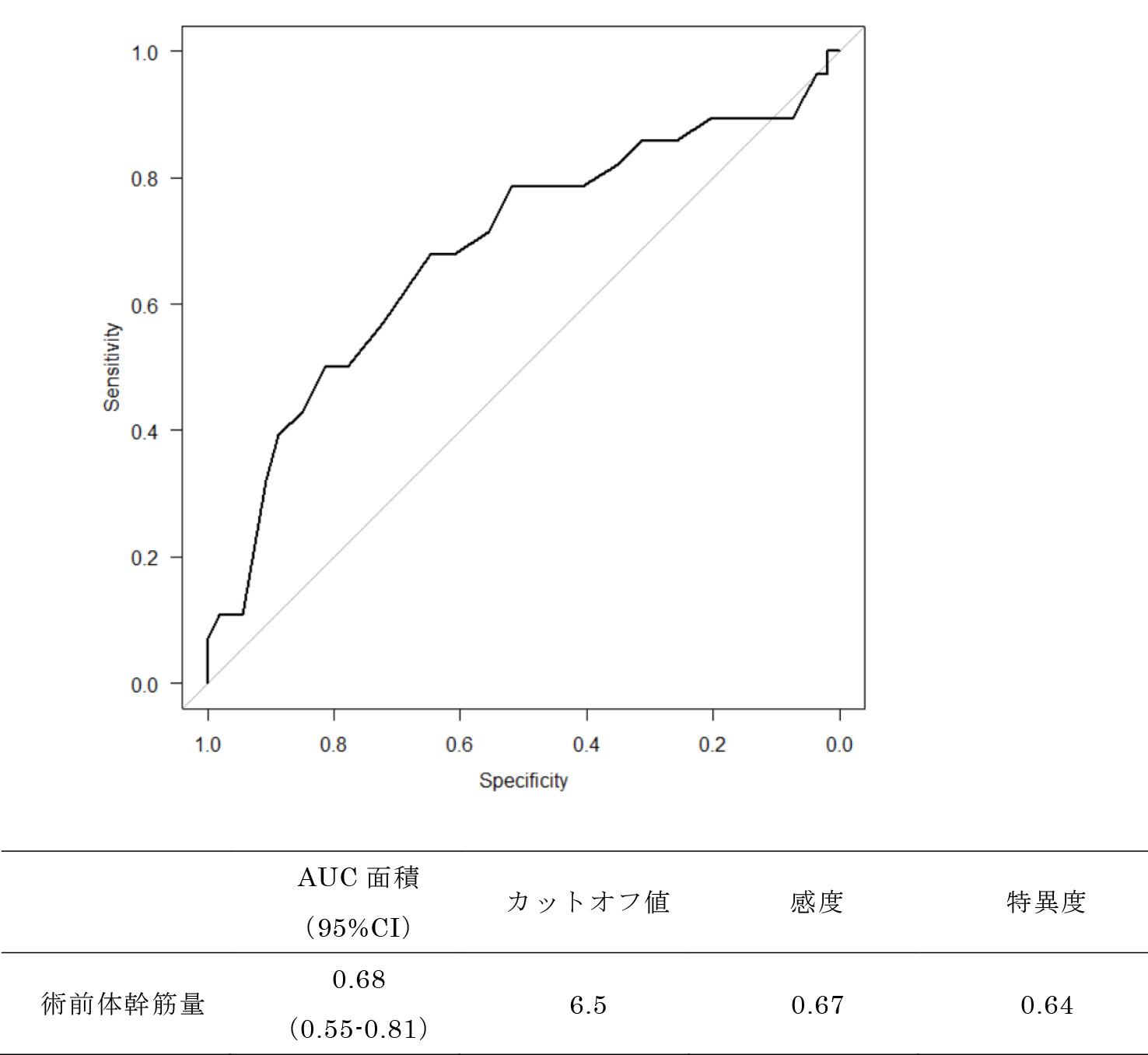
    Methods: Eighty-two patients who underwent spinal corrective fusion for adult spinal deformity were included (mean age 73.6 years). Preoperative trunk muscle mass was calculated from bioimpedance analysis. Postoperative back pain was defined as back pain of 30 mm or more on a visual analog scale. In the statistical analysis, cutoff values for preoperative trunk muscle mass were calculated using receiver-operating characteristic (ROC) curves, with the presence or absence of postoperative back pain as the anchor. Next, univariate and multivariate adjusted models were analyzed to examine whether the calculated cutoff values could predict the presence or absence of postoperative back pain (significance level 5%).

    Results: The ROC curve showed that the cutoff value for preoperative trunk muscle mass was 6.5 kg/m2 (area under the ROC curve 0.68, sensitivity 0.67, specificity 0.64). Univariate and multivariate adjusted models showed that the cutoff value of trunk muscle mass was a predictor of the presence or absence of postoperative back pain in all models.

    Conclusions: The calculated cutoff value of preoperative trunk muscle mass is moderately accurate and can be used to predict the presence or absence of low back pain in patients undergoing spinal corrective surgery.

    Download PDF (951K)
  • Masashi Urayama, Yuko Takeda, Hidetaka Furuya, Masahiro Hoshino
    2025 Volume 16 Issue 6 Pages 873-879
    Published: June 20, 2025
    Released on J-STAGE: June 20, 2025
    JOURNAL FREE ACCESS
    Supplementary material

    Introduction: Cases in which surgery was performed for patients aged ≥90 years with osteoporotic vertebral fractures were investigated.

    Methods: A total of 23 vertebrae of 22 patients aged ≥90 years or older with osteoporotic vertebral fractures (OVFs) who underwent surgical treatment at our hospital between 2011 and 2023 were retrospectively investigated. The investigation included age, sex, medical history, fractured vertebrae, preoperative bone density, preoperative waiting period, surgical procedures, surgical duration, length of hospital stay, and mobility at discharge.

    Results: The average age was 91.8 years, with 6 males and 16 females. The most common pre-existing condition was hypertension, and the most frequently fractured vertebra was the first lumbar vertebra. The mean young adult mean (YAM) value was 62.14. The preoperative waiting period ranged from a minimum of 5 days to over one year. The surgical procedures included balloon kyphoplasty (BKP) alone, vertebral body stenting (VBS) alone, and a combination of BKP and pedicle screw fixation (PPS). The average surgery time was 24.7 minutes for BKP or VBS alone and 104.3 minutes for the combination of BKP and PPS. The average hospital stay was 29.6 days. At discharge, mobility outcomes were as follows: 11 patients used wheelchairs, 5 used walkers, 3 were ambulatory without assistance, and 3 used canes. There were no cases of severe postoperative complications.

    Conclusions: Complications and decreaced activities of daily living due to OVF in patients aged ≥90 years are unavoidable. Although the usefulness of surgeries such as BKP has been reported, the hurdles for performing such procedures are not low.

    Download PDF (1708K)
  • Masayuki Ishihara, Shinichiro Taniguchi, Masaaki Paku, Yoichi Tani, Ta ...
    2025 Volume 16 Issue 6 Pages 880-890
    Published: June 20, 2025
    Released on J-STAGE: June 20, 2025
    JOURNAL FREE ACCESS

    Introduction: We investigated the risk factors of poor outcomes in MIS short fusion for adult spinal deformity (ASD).

    Subjects and Methods: The study included 82 patients with adult spinal deformity (ASD) who had undergone MIS-short fusion using MIS techniques such as LLIF, ACR, and PPS at T12 or below, and who had been able to be followed up for at least 2 years. The average age was 73.7 years, and the average follow-up period was 35.5 months. The definition of poor results was final SVA > 50 mm or the development of PJF/DJF. Firstly, we divided the patients into Group G (final SVA≤50 mm) and Group P (final SVA>50 mm), and examined the femoral bone mineral density, number of fixation ranges, HU value (UIV), various parameters, GAP score, ODI, and walking speed. Next, we examined the same items in two groups: Group JF (with JF) and Group NJF (without JF).

    Results: In the first analysis, there were no significant differences in age, gender, bone mineral density, or HU value, and the fixed range was significantly shorter in the G group. In the G group, the preoperative and postoperative LL were significantly larger, the PI-LL was significantly smaller, and the preoperative and postoperative SVA were significantly smaller. In addition, there were significant differences in the RLL (LL-ideal LL) and RSA (global tilt (GT) -ideal GT) in the GAP score. There was no significant difference in gait speed, and the postoperative ODI was significantly smaller in Group G. In multivariate logistic regression analysis with the final-SVA>50 mm as the objective variable, only the preoperative SVA was detected as a risk factor. Similarly, univariate and multivariate analyses were performed on the JF and NJF groups, and the risk factors for JF were found to be GAP score.

    Conclusion: In MIS short fusion for ASD, preoperative SVA is important for final SVA, and the importance of GAP score in the occurrence of JF was suggested.

    Download PDF (1897K)
  • Kiyonori Yo, Yoko Matsuda, Yosuke Oishi, Eiki Tsushima, Masaaki Murase ...
    2025 Volume 16 Issue 6 Pages 891-899
    Published: June 20, 2025
    Released on J-STAGE: June 20, 2025
    JOURNAL FREE ACCESS

    Introduction: The McKenzie method (mechanical diagnosis and therapy [MDT]) is an exercise treatment to identify appropriate exercises and postures that improve symptoms and then apply them. This study's aim was to investigate whether MDT was effective for sub-acute low back pain and to analyze factors related to its effectiveness.

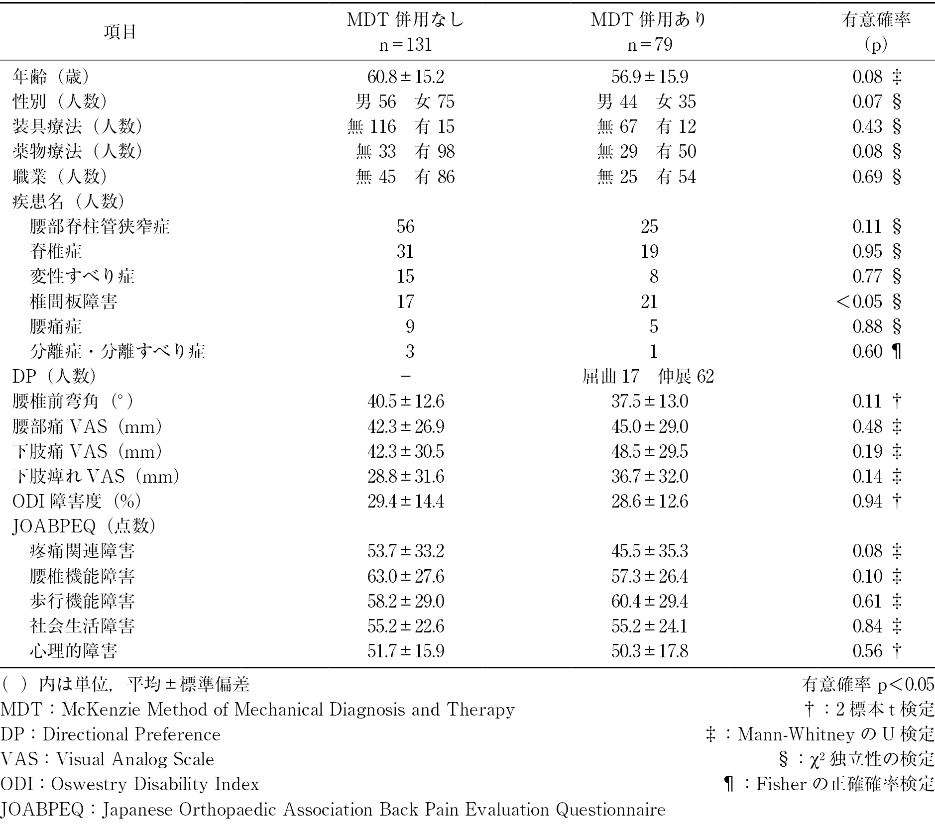
    Methods: This was a retrospective, cohort study involving 210 patients who had low back pain for >4 weeks and <3 months and received outpatient treatment at our hospital. The participants were divided into two groups according to whether they were treated with MDT (MDT [+], n = 79) or not (MDT [−], n = 131). All patients performed stretching and trunk muscle exercises. Clinical results were compared between the two groups after 1 and 3 months of treatment using a visual analog scale (VAS), Oswestry Disability Index (ODI), and Japanese Orthopaedic Association Back Pain Evaluation Questionnaire (JOABPEQ). Factors related to the effectiveness of MDT were also analyzed.

    Results: Each clinical measure was higher after 1 and 3 months of treatment than before in both groups. The MDT (+) group had a significantly higher increase in the score for pain-related disability and a higher efficacy rate for pain-related disability and lumbar dysfunction of the JOABPEQ than the MDT (−) group. The group with the effect of MDT had more severe lumbar dysfunction before treatment than the group without.

    Conclusions: In patients with sub-acute low back pain, the MDT (+) group showed significantly better clinical outcomes than the MDT (−) group. This suggested that MDT might be a useful exercise treatment for sub-acute low back pain.

    Download PDF (1406K)
  • Toshihiro Imamura, Tomohiko Uemori, Masato Yoshimoto, Hiroki Tanaka
    2025 Volume 16 Issue 6 Pages 900-904
    Published: June 20, 2025
    Released on J-STAGE: June 20, 2025
    JOURNAL FREE ACCESS

    Introduction: There are few reports on return to work after intradiscal enzyme injection therapy (Condoliase) and Microendoscopic Discectomy (MED) for lumbar disc herniation. This study aimed to compare the return-to-work timing between patients who underwent Condoliase and MED and to examine the impact of patient background factors (such as age and smoking status) on return to work.

    Methods: From September 2019 to March 2024, 37 working patients who underwent Condoliase or MED at our institution were included, excluding three patients who required reoperation. Patient age, sex, occupation, smoking status, return-to-work timing, and return-to-work status were analyzed.

    Results: The Condoliase group included 13 patients (average age 51.5 years; 10 males, 3 females), with 33.3% being smokers. The average time to return to work after surgery was 12.9 days. The MED group included 24 patients (average age 44.0 years; 18 males, 6 females), with 66.7% being smokers. The median time to return to work was 14.0 days, with an average of 16.4 days. Both groups returned to their preoperative occupations.

    Discussion: Although we hypothesized that the return-to-work period would be shorter for the Condoliase group, no significant difference was observed (Condoliase: mean 12.9 days; MED: mean 16.4 days). Early pain relief after Condoliase treatment did not necessarily lead to a quicker return to work, likely due to patient or employer concerns regarding post-surgical recovery. Additionally, some patients were instructed by their workplaces to remain on leave for up to one month postoperatively. Smokers tended to choose MED, seeking a more definitive treatment, while non-smokers prioritized minimally invasive treatment and opted for MED if the effect was insufficient. Due to the impact of the COVID-19 pandemic, there was an increase in patients preferring short-term hospitalization, and some cases showed a more cautious return to work.

    Conclusion: Both Condoliase and MED resulted in a median return-to-work period of 14 days. Statistical analysis indicated a significant association between smoking status and surgical selection (p < 0.05), with smokers being more likely to choose MED.

    Download PDF (949K)
  • Shuhei Iwata, Kenji Hatakeyama, Atsushi Kojima, Shigeru Kamitani, Naok ...
    2025 Volume 16 Issue 6 Pages 905-911
    Published: June 20, 2025
    Released on J-STAGE: June 20, 2025
    JOURNAL FREE ACCESS

    Introduction: Lumbar spondylolysis is a fatigue fracture of the pars interarticularis. When diagnosed early, bone healing can often be achieved through bracing. However, some cases fail to heal in patients unable to use bracing or because they are already in an advanced stage at the time of diagnosis, making union difficult. Given that extracorporeal shock wave therapy (ESWT) has shown bone-healing effects in fresh fractures and pseudarthrosis of long bones, we hypothesized that it may be beneficial for lumbar spondylolysis as well. This study investigated the safety and efficacy of ESWT for patients with lumbar spondylolysis.

    Methods: We included 46 patients (64 lesions) diagnosed with lumbar spondylolysis between January 2022 and June 2024 at our institution, all of whom consented to participate and were available for follow-up. Treatment was conducted using the DUOLITH (STORZ MEDICAL) system, with fluoroscopy marking the lesion site, and shock waves were applied biweekly to painful areas. Intensity was adjusted to the tolerable range of 0.15-0.25 mJ/mm2, with 3,000 pulses per session. We assessed complication rates, bone union rates, and time to bone union.

    Results: No complications, such as nerve damage or ligament ossification, were observed. The overall bone union rate was 78%, with an average time to union of 2.5 months. By disease stage, union rates and times were as follows: 100% union in 1.7 months for 18 cases in the earliest stage; 100% union in 2 months for 15 cases in the early stage; 76% union in 3 months for 16 cases in the progressive stage; and 10% union in 12 months for 10 cases in the end stage.

    Conclusions: Despite including 22 patients who underwent ESWT without bracing to continue sports activities, the bone union rate and time to union were both improved compared to traditional methods. ESWT provides a promising, non-invasive treatment option for lumbar spondylolysis, offering high union rates and expedited healing times.

    Download PDF (1903K)
  • Yoko Matsuda, Eiki Tsushima, Kiyonori Yo, Yosuke Oishi, Masaaki Murase
    2025 Volume 16 Issue 6 Pages 912-918
    Published: June 20, 2025
    Released on J-STAGE: June 20, 2025
    JOURNAL FREE ACCESS

    Introduction: The purpose of this study was to investigate whether the time of postpartum low back pain onset affected the clinical outcomes of physical therapy for low back pain.

    Subjects and Methods: A total of 50 patients participated in this study; 18 had onset of low back pain less than one year after childbirth (early group). and 32 had onset after more than one year (late group). All patients received the same exercise therapy and lifestyle guidance. Clinical results were compared between the two groups after 1 and 3 months of treatment using a visual analog scale (VAS) for low back pain and each item and the total of the Oswestry Disability Index (ODI).

    Results: In both groups, Sitting, Standing, Sleeping, and Travel of the ODI and the total ODI improved significantly 1-3 months post-treatment from pre-treatment. However, no significant differences were found between the groups. The VAS for low back pain and, Pain intensity, Personal care, Lifting, Walking, and Social of the ODI improved significantly 1-3 months post treatment from pre-treatment in the late group, but improved significantly 3 months post treatment from pre-treatment in the early group.

    Conclusion: In the course of physical therapy for patients with postpartum low back pain, both groups showed improvements in static daily living functions, while the late group demonstrated improvements in dynamic daily living functions from as early as one month. Physical therapy may be effective for patients with postpartum low back pain.

    Download PDF (1393K)
  • Masatoshi Inoue, Hidehisa Torikai, Yoshinori Nakata
    2025 Volume 16 Issue 6 Pages 919-926
    Published: June 20, 2025
    Released on J-STAGE: June 20, 2025
    JOURNAL FREE ACCESS

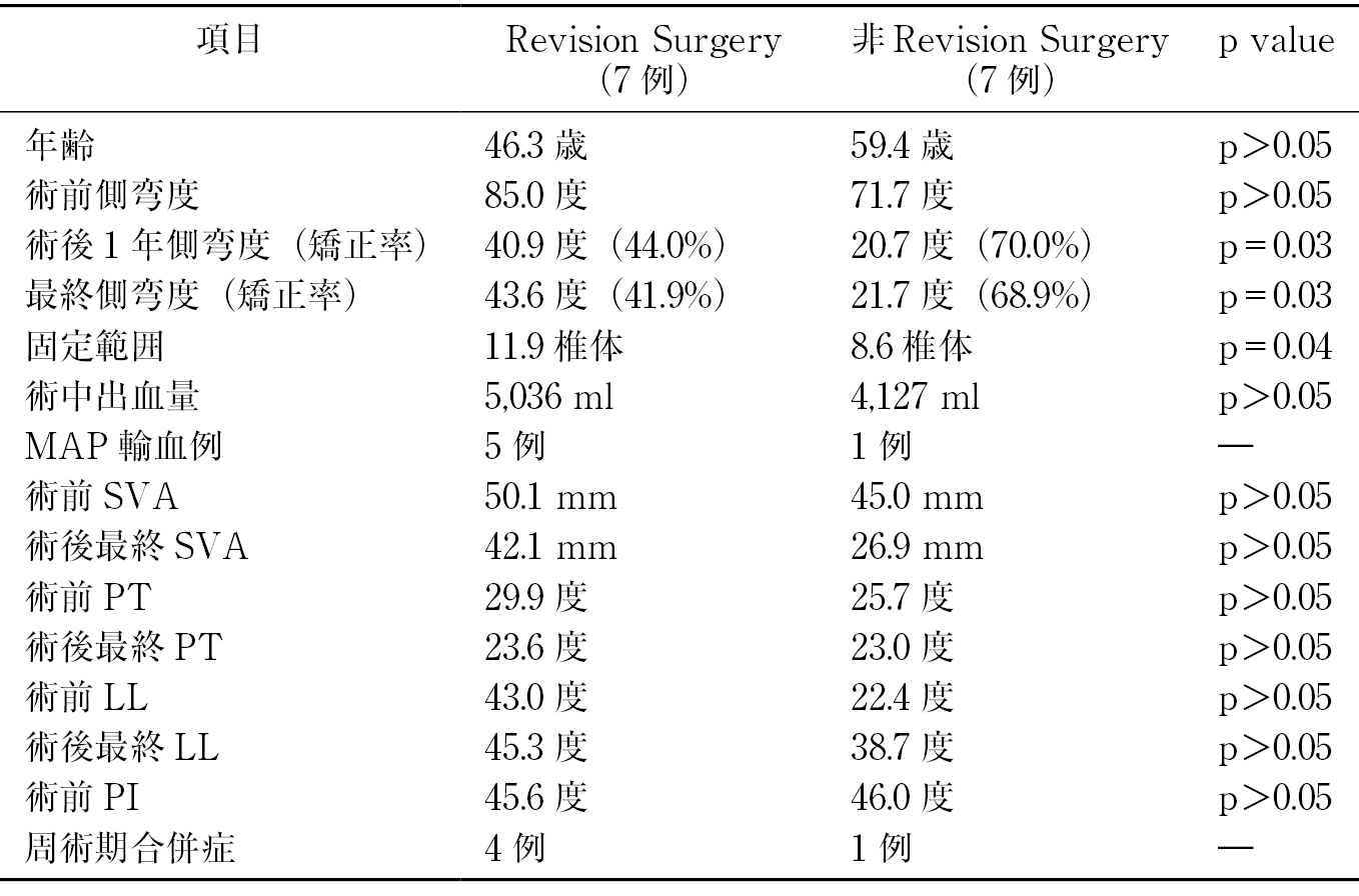
    Introduction: Attention to adult spinal deformity (ASD) has increased over the past few decades, resulting in important clinical advances in its treatment. Three-column osteotomy, including vertebral column resection (VCR), is a powerful method to correct rigid scoliosis. The purpose of this study was to evaluate surgical treatment with VCR.

    Patients and Methods: Fourteen patients (3 men and 11 women; mean age 56.2 years) with severe ASD were treated surgically. The mean preoperative Cobb angle was 71.8°. The preoperative diagnoses included revision scoliosis surgery due to curve progression in seven patients, ASD with known scoliosis in adolescence in four patients, de novo ASD in two patients, and congenital kyphosis in one patient. All patients complained of back pain, one had spinal cord injury with complete paraplegia, and two patients had muscle spasticity and difficulty walking. According to the ASD SRS-Schwab classification, seven patients were classified with marked sagittal imbalance (SVA>9.5 cm, PT>30°, PI-LL>20°).

    Results: Thirteen of 14 patients underwent single-stage posterior VCR. One patient had two-stage VCR. The level of the resected vertebra was L1 in seven patients, T12 in two patients, L2 in two patients, and T8, 9, or 11 in one patient each. The mean postoperative Cobb angle was 30.8°, corresponding to a mean correction rate of 57.2%. In two patients, marked sagittal imbalance remained. The mean preoperative blood transfusion was 1,800 ml, and the mean estimated intraoperative blood loss was 5,129 ml. Six patients required homologous blood transfusions, five patients had surgical complications, two had respiratory dysfunction, two had temporary neurological complications, and one patient developed a surgical site infection.

    Conclusion: Although the rate of complications was high, VCR surgery for patients with ASD can be used effectively even for revision scoliosis surgery. The routine use of high-dose tranexamic acid might reduce blood loss.

    Download PDF (1550K)
Case Report
  • Naoki Tsujishima, Shigeru Kamitani, Atsushi Kojima, Hirohito Suzuki, T ...
    2025 Volume 16 Issue 6 Pages 927-931
    Published: June 20, 2025
    Released on J-STAGE: June 20, 2025
    JOURNAL FREE ACCESS

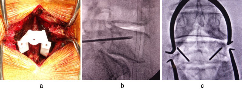
    Introduction: The fixation strength of cortical bone trajectory (CBT) pedicle screws in spondylolytic vertebrae has been reported to be limited. A long CBT, also known as the midline cortical (MC) trajectory, is expected to provide increased fixation strength when inserted deeper than 40% of the vertebral body length. Patient-specific guides have been considered technically difficult to apply to a spondylolytic pars; however, a case of successful MC screw placement using a patient-specific guide is reported.

    Case Report: A 76-year-old man presented with right leg pain and intermittent claudication. Imaging showed L5 bilateral spondylolysis with Grade 1 slip (Meyerding classification), and L4/5 laminectomy and L5/S transforaminal lumbar interbody fusion were performed. A patient-specific guide (MySpine MC®) was placed on the spondylolytic pars, and MC screws were safely inserted under fluoroscopic guidance. At 6-month follow-up, there was no evidence of screw loosening or displacement, and neurological symptoms had resolved completely.

    Conclusions: Safe MC screw insertion was performed using a patient-specific guide in lumbar spondylolysis, achieving good clinical outcomes. This technique suggests that MC screws can be safely and accurately inserted using a patient-specific guide even in the presence of spondylolytic pars. Further studies with larger patient cohorts are needed to validate these findings.

    Download PDF (1497K)
Secondary Publication
  • Takaya Imai, Sota Nagai, Takehiro Michikawa, Risa Tobe, Soya Kawabata, ...
    2025 Volume 16 Issue 6 Pages 932-938
    Published: June 20, 2025
    Released on J-STAGE: June 20, 2025
    JOURNAL FREE ACCESS

    Introduction: The treatment of patients with lumbar spinal canal stenosis (LSCS) is mainly divided into conservative and surgical treatments, but conservative treatment, including drug therapy, is the first choice. It is expected that the dependence of LSCS patients who have undergone surgical treatment on conservative treatment will decrease, and the use of drugs will decrease. The purpose of this study is to examine how surgical treatment affects drug therapy in LSCS patients and to identify the factors associated with an increase in the use of drugs after surgery.

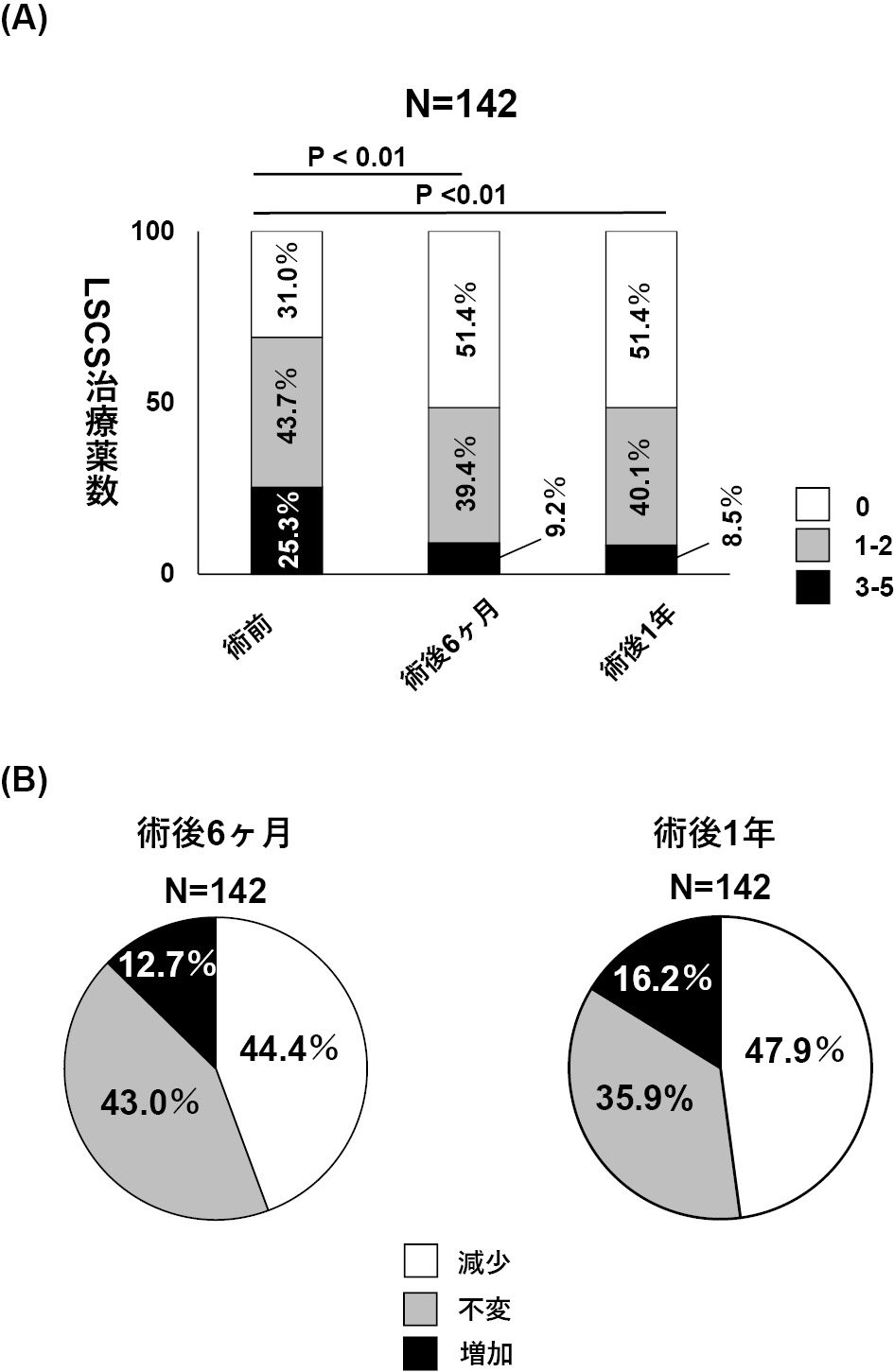
    Materials: The subjects were 142 patients (84 men and 58 women, average age 70.1 years) aged 40 years or older who underwent lumbar surgery for LSCS at our hospital between April 2020 and March 2021. We retrospectively examined information from before and 6 months and 1 year after surgery, and evaluated patient background, patient-reported questionnaires including JOABPEQ, and postoperative prescription drugs.

    Results: Lumbar surgery for LSCS patients overall significantly reduced the number of LSCS medications, but the number of medications increased in approximately 15% of patients. Poor postoperative outcomes for walking ability and social life in JOABPEQ were significantly associated with an increase in the number of LSCS medications after surgery.

    Conclusion: Lumbar surgery for LSCS patients reduced the number of LSCS medications, but poor postoperative outcomes for walking ability and social life were potentially involved in the increase of the number of medications.

    Download PDF (1235K)
feedback
Top