Progress of Digestive Endoscopy
Online ISSN : 2187-4999
Print ISSN : 1348-9844
ISSN-L : 1348-9844
Volume 58, Issue 2
Displaying 1-44 of 44 articles from this issue
Technology and instrument
  • Hiroyuki Kato, Shungo Endo, Takao Katsube, Shunsuke Haga, Tetsuro Kaji ...
    2001 Volume 58 Issue 2 Pages 24-26
    Published: June 15, 2001
    Released on J-STAGE: May 31, 2014
    JOURNAL FREE ACCESS
    The new colonoscopes of which the shaft is stiffness-ajustable are evaluated in relation to the performance of insertion on total colonoscopy. In order to evaluate practical usefulness, 282 cases were examined with either adjustable scopes (group A) or usual scopes as control (group B) . When we use adjustable scopes, the examination was started in the most soft state of the shaft, and the stiffness was changed to the most hard state when the scope was not able to insert to deeper portion due to the bending of the scope. If the adjustment of the stiffness was not effective, we use the abdominal manipulation. Evaluated items are insertion rate and time to the cecum, the degree of the patient's pain, the rate of using the abdominal manipulation in order to protect the bending of the scope. The adjustment of stiffness (from soft to hard) of the shaft was performed in 51% of group A. No significant difference was noted between two groups in terms of insertion rate (98% in group A vs 99% in group B) , and time (5'02” vs 5'15”) to the cecum and the degree of the pain. In group A, the rate of using the abdominal manipulation is 31%, that is significantly low in relation to 67% in group B.
    In conclusion adjustable scopes could reduce the effort of the assistant because of decreasing the rate of using the abdominal manipulation, and it was recognized that those scopes are useful on the insertion to the deep colon.
    Download PDF (333K)
Clinical study
  • Yuhsaku Kanoh, Tadashi Ohara, Isao Okayasu
    2001 Volume 58 Issue 2 Pages 28-32
    Published: June 15, 2001
    Released on J-STAGE: May 31, 2014
    JOURNAL FREE ACCESS
    The effects of various factors in regeneration of gastric mucosa in patients who have chronic renal failure (CRF) and have been treated with hemodialysis (HD) , were studied. The gastric mucosa obtained by endoscopy was immunohistologically stained. The CRF patients were divided into 3 groups according to the length of HD therapy. Group I included those who had received HD for more than 4 years (n=25) , Group II included those who had received HD for less than 4 years (n=20) , and Group III included those who had never been treated with HD (n=13) . On immunohistological studies, their speciments of gastric mucosa gave positive stain for ki-67. The positive labeling index (LI) of ki-67 in the HD group was greater than 80%, while it was 20.6% in non-HD group. However, there was no significant differences in mucosa-regenerating factors[gastrin, cholecystokinin (CCK) and epidermal growth factor (EGF) ]between the HD group and non-HD group. On the other hand, the percentage of patients in the HD group who were infected with Helicobacter pylori was lower than that in the non-HD group. These results suggest that HD therapy in CRF patients induces regeneration of the gastric mucosa, probably by removing those factors that contribute to atrophy of the gastric mucosa.
    Download PDF (1510K)
  • Toshiro Kamoshida, Soichi Hotta, Shinji Hirai, Kazuto Takamura, Tsunet ...
    2001 Volume 58 Issue 2 Pages 34-38
    Published: June 15, 2001
    Released on J-STAGE: May 31, 2014
    JOURNAL FREE ACCESS
    Foveolar hyperplastic polyps are the most popular polyp of the stomach arise from the mucosa of chronic active gastritis. Recent studies suggested hyperplastic polyps arise from inflammatory process of H. pylori gastritis and disappeared or reduced after H. pylori eradication therapy. We report four cases of foveolar hyperplastic polyps of the stomach disappeared and reduced after H. pylori eradication therapy. Including a case of Group IV lesion (adenocarcinoma suspected polyp) disappeared after H. pylori eradication therapy. In all cases, foveolar hyperplastic polyps of the stomach disappeared and reduced after H. pylori eradication therapy. Group IV lesion (adenocarcinoma suspected polyp) also disappeared after H. pylori eradication therapy confirmed by endoscopic mucosal resection. Histological examination of the polyps compared before and after H. pylori eradication therapy revealed foveolar epithlial hyperplasia, inflammatory cell infiltration and stromal edema decreased after H. pylori eradication therapy. Accompany with that, MIB-1 (Ki-67) Labelling index, proliferating cell marker, also decreased markedly. A case of Group IV lesion (adenocarcinoma suspected polyp) disappeared after H. pylori eradication therapy was very rare. It suggested adenocarcinoma arise from hyperplastic polyp confirmed by immunostaining of p53 protein may disappear by H. pylori eradication therapy.
    Download PDF (1309K)
  • Ken-ichi Nakajima, Taketo Yamaguchi, Tateo Odaka, Noriko Hayashida, Sh ...
    2001 Volume 58 Issue 2 Pages 40-43
    Published: June 15, 2001
    Released on J-STAGE: May 31, 2014
    JOURNAL FREE ACCESS
    Between 1994 and 1999, we had experienced 17 cases of gastric anisakiasis at Tako Central Hospital, Which is located in the north eastern part of Chiba Prefecture. Seven patients (41.2%) developed gastric anisakiasis after eating raw sardines and 2 (11.8%) after eating raw bonito. The disease was prevalent between December and May, i.e., the period when sardines abound. Fourteen patients (82.4%) developed symptoms at night and upper abdominal pain was the commonest symptom (16 patients) . Twelve patients (70.6%) visited the hospital within 12 hours since the onset of symptoms. However, three patients waited 2 days or longer to see the doctor. Gastric anisakiasis usually presented as upper abdominal pain of sudden onset and anamnesis was most important for a correct diagnosis of the infection because it does not follow a typical pattern. As for the number of Anisakis larvae found at the gastroscopy, there was one larva in 15 patients (88.2%) , two larvae in one and four larvae in one patient. The gastric mucosa at the site of the larvae penetrated the tissues most frequently presented edema, erosion and redness. The mucosa often showed erosion, spotty coagulation and superficial gastritis in other areas. These results indicate that close gastroscopic examination of the gastric mucosa, especially at the corpus, should be done when a patient is suspected of having anisakiasis.
    Download PDF (474K)
  • Hidejiro Urakami, Kouichi Aiura, Yoshio Matsui, Yasushi Iwasaki, Kouic ...
    2001 Volume 58 Issue 2 Pages 44-48
    Published: June 15, 2001
    Released on J-STAGE: May 31, 2014
    JOURNAL FREE ACCESS
    There are recently various approaches for the treatment of common bile duct stones (CBDS) including open surgery, laparoscopic surgery, and endoscopic procedures.
    In our institution, we usually perform endoscopic papillary balloon dilation (EPBD) with precut sphincterotomy (preEST) for CBDS more than 1 cm in diameter as our policy.
    From November in 1996 to August in 2000, we treated 44 cases with CBDS by EPBD and 45 cases by EPBD combined with preEST. The average period of observation was 23.6 months. Recurrent CBDS were observed in 3 cases. Two cases were the patients who retained the gallbladder (GB) with stones after EPBD. The cystic ducts were slightly dilated and the gallbladders were not visualized at the initial cholangiography in these cases. This finding suggests that GB stones could be easy to move to CBD after EPBD in the patients with so-called negative GB at the initial cholangiography. Rest of one case underwent laparoscopic cholecystectomy after EPBD combined with EST, however, recurrent CBDS was observed one year and 9 months later. This patient had received hemodialysis for chronic renal failure. Although there are controversial opinions on the prevalence rate of cholelithiasis in patients undergoing dialysis, it is suggested that we need to take care of these patients carefully for the reccurence of CBDS.
    Download PDF (3287K)
  • Terumi Kamisawa, Yuyang Tu, Naoto Egawa, Nobuhiro Sakaki, Jun-ichi Ish ...
    2001 Volume 58 Issue 2 Pages 50-54
    Published: June 15, 2001
    Released on J-STAGE: May 31, 2014
    JOURNAL FREE ACCESS
    Choledochocele was first described as a cystic dilatation of the intramural segment of the common bile duct protruding into the duodenal lumen in 1940. However, no definitions in detail have been discussed and the etiology remained unclear. We studied retrospectively 5,500 cases of ERCP and discussed about analogous cystic diseases to choledochocele as follows ; cases with a dilated the common channel whose diameter was under 1 cm (dilated common channel syndrome) , diverticular protrusion of the distal common bile duct whose diameter was as small as 5 mm, dilatation of opening of the main pancreatic duct (pancreatocele) , associated with choledocholithiasis or after stone extraction, associated with pancreaticobiliary maljunction. Criteria of choledochocele should be made including the minimum diameter, congenital or acquired, concepts of dilated common channel and pancreatocele, and relationship to pancreaticobiliary maljunction.
    Download PDF (3354K)
Case report
  • Osamu Kawamura, Motoyasu Kusano, Keiko Minashi, Masaki Maeda, Sayaka T ...
    2001 Volume 58 Issue 2 Pages 56-59
    Published: June 15, 2001
    Released on J-STAGE: May 31, 2014
    JOURNAL FREE ACCESS
    We experienced two cases of gastrointestinal stromal tumor (GIST) with definite growth. Both tumors showed a homogeneous internal echogenecity. EUS-guided aspiration needle biopsy revealed few mitotic figures and leiomyoma was diagnosed by H-E stain. Because both tumors showing growth, partial gastrectomy was performed via laparoscopy. Histopathological examination yielded a diagnosis of GIST (uncommitted type) using stain for c-kit and other immunohistochemical markers. Since 1-2 mitotic figures were seen per 10 HPF, malignancy was suggested. Therefore, it is necessary to give careful consideration to malignancy if the tumor is growing more than 7 to 8 mm per one year, even though there is little evidence of a malignant lesion on EUS or biopsy. Also, biopsy tissue should be examined using immunohistochemical stain to diagnose GIST. Even if there are few mitotic figures, consideration must be given to the degree of biological malignancy of GIST when evaluating the indications for surgery.
    Download PDF (2001K)
  • Yoh Isobe, Kiyoshi Kubochi, Atsushi Shimada, Hiroya Takeuchi, Takashi ...
    2001 Volume 58 Issue 2 Pages 60-63
    Published: June 15, 2001
    Released on J-STAGE: May 31, 2014
    JOURNAL FREE ACCESS
    Case 1 was a 66-year-old male who had developed a type IIa cancer in the posterior wall of the remnant stomach (RS) 69 months after a distal gastrectomy for a type 2 cancer in the lower stomach. The histological type of the primary cancer was a moderately differentiated adenocarcinoma. Severe bile reflux was observed in the RS. Helicobacter Pylori (HP) was negative. Case 2 was a 33-year-old male who had developed a type IIa cancer near the suture line of the RS 8 months after a distal gastrectomy for a type IIa+IIc cancer in the middle stomach. The histological type of the primary cancer was a well differentiated adenocarcinoma. He had a synchronous colon cancer. HP was positive. Case 3 was a 51-year-old male who had developed a type IIc cancer in the anterior wall of the RS 25 months after a laparoscopic partial gastrectomy for a type IIc cancer in the middle stomach. The histological type of the primary cancer was a well differentiated adenocarcinoma. HP was positive. The RS cancers were detected by the postoperative endoscopic examination that had been carried out once a year in case 1 and twice a year in case 3. All of the lesions in 3 cases could be treated by EMR. No recurrent lesions have been observed for 18, 14 and 10 months after EMR in cases 1, 2 and 3 respectively.
    To treat the RS cancer successfully by EMR, postoperative intensive surveillance for the high-risk patients might be necessary.
    Download PDF (432K)
  • Takeshi Shimizu, Yoshihisa Kuzutani, Kouichi Kuroda, Moriyoshi Tanaka, ...
    2001 Volume 58 Issue 2 Pages 64-67
    Published: June 15, 2001
    Released on J-STAGE: May 31, 2014
    JOURNAL FREE ACCESS
    Paneth cells are identified by their distinctively large eosinophilic cytoplasmic granules normally found in the Lieberkuhn's gland. It is also commonly known that they appear in the intestinal metaplastia of the stomach. But recently there are some reports of paneth cells found in the gastric carcinomas which is still not widely known.
    A 67-year-old male patient was diagnosed as early gastric carcinoma by routine endoscopic examination performed in July, 2000. The gastric carcinoma was histopathologically diagnosed as well differentiated adenocarcinoma. Paneth-like tumor cells were found in the gastric carcinoma membrane and was distinctively identified by PTAH and other staining and also by the existence of lysosome. And further investigation revealed that Ki-67 and PCNA was both positively stained in the tumor cells and p-53 was negatively stained.
    Domestically gastric carcinoma with paneth-like tumor cells have been reported since 1966 by Sano and since then 40 cases have been reported. A precise study of these cases revealed that 0.3% to 4.1% of the gastric carcinoma were associated with paneth-like tumor cells. Among these 40 cases, 28 cases were early carcinoma against 9 cases of advanced carcinoma. Also classified by their histological type, 29 cases were well differentiated type against only 6 cases of undifferentiated type. Surrounding these carcinoma, intestinal metaplasia were found in 34 cases. These results suggest that gastric carcinoma with paneth-like tumor cells were likely to be found in the early stage of well differentiated carcinoma and has strong relationship to intestinal metaplasia itself. These facts might give a new lead to the histogenesis of well differentiated type gastric carcinoma.
    Download PDF (1095K)
  • Yoshinori Hayashi, Toshiaki Sugiura, Makoto Kotoyori, Hisagi Ishikawa, ...
    2001 Volume 58 Issue 2 Pages 70-71
    Published: June 15, 2001
    Released on J-STAGE: May 31, 2014
    JOURNAL FREE ACCESS
    We report here an 83-year-old woman with an early esophageal adenocarcinoma arising from Barrett's esophagus. Endoscopic examination revealed a reddish depressive lesion 3 mm in diameter and a nodular-faced lesion in columnar epithelium. Biopsy specimen from both lesions revealed well-differentiated adenocarcinoma.
    We performed subtotal esophagectomy and the resected specimen showed the length of Barrett's esophagus to be about 7 cm. The type 0-IIc esophageal carcinoma measured 3 mm and the type 0-IIa esophageal carcinoma measured 1 cm in size. Well-differentiated adenocarcinoma was presented in both lesion, and in both, the invasion depth was limited to the lamina propria mucosa. There was no evidence of lymph node metastasis. Barrett's esophagus is considered a precancerous condition and the treatment of choice with curative intent has been esophagectomy. Subtotal esophagectomy was performed safely for this 83-year-old patient and we suggest that subtotal esophagectomy is an effective therapy for early adenocarcinoma in Barrett's esophagus.
    Download PDF (213K)
  • Ueki Hisayo, Nobuhiko Hatano, Atushi Chiba, Yoshitake Ikeda, Yutaka At ...
    2001 Volume 58 Issue 2 Pages 72-73
    Published: June 15, 2001
    Released on J-STAGE: May 31, 2014
    JOURNAL FREE ACCESS
    A 70-year-old male was admitted to our hospital because of hematoemesis and tarry stool. Endoscopic findings showed semicircle esophageal cancer which measuring about 5cm between upper and middle thoracic esophagus, and then, he was diagnosed for the first time as achalasia. Surgery for thoracic esophageal cancer was performed. Pathological examination of resected specimen revealed squamous cell carcinoma, moderately differentiated type, of esophagus, and depth of invasion mp.
    In this case, the patient had no symptom. However after operation he found having dysphagia before. Achalasia is known to high risk of esophageal cancer. We should find and follow up esophageal achalasia patients with esophageal cancer in mind.
    Download PDF (1433K)
  • Katsuhiko Higuchi, Masato Ooida, Satoshi Tanabe, Mitsuhiro Kida, Wasab ...
    2001 Volume 58 Issue 2 Pages 74-75
    Published: June 15, 2001
    Released on J-STAGE: May 31, 2014
    JOURNAL FREE ACCESS
    A 62-year-old man had a medical examination on our hospital on March 7, 2000. He had complained of dysphagia for a year. Esophagogram revealed multiple flask-shaped outpouchings in the upper intrathoratic esophagus, then he was diagnosed as esophageal intramural pseudodiverticulosis (EIPD) . Esophagoscopy on April 10, 2000, showed multiple white plaques and mild stenosis in the upper intrathoratic esophagus. Histological examination demonstrated inflammatory cells which massively infiltrated into the epithelium, and presence of Candida Albicans. Therefore, he was diagnosed as EIPD with Candida esophagitis. After antifungal medications were administered for a week, dysphagia was improved. Esophagoscopy on May 9, 2000, revealed multiple orifices of the pseudodiverticula. We reviewed Esophagogram from another hospital, then we found that EIPD had grown up for a year.
    Download PDF (1193K)
  • Masayuki Sekine, Kimiaki Hattori, Takayuki Amano, Yoshimasa Suzuki, Na ...
    2001 Volume 58 Issue 2 Pages 76-77
    Published: June 15, 2001
    Released on J-STAGE: May 31, 2014
    JOURNAL FREE ACCESS
    A 22-year-old male sufferd upper aerodigestive tract burn by an unexpected explosion of firework in his mouth. A month later, esophageal stricture developed and needed weekly endoscopic dilatation. 7 months later, he was introduced to our hospital for treatment of his esophageal stricture. He could not swallow down meat and fish. Endoscopic examination revealed esophageal stenosis through 18cm to 22cm from incisor, and a narrow diameter endoscope (8mm in diameter) could not pass through the stenotic lesion. After treatment with a balloon dilator (12mm in diameter) , a panendoscope (11mm in diameter) could pass through the stenotic lesion. Then, he underwent a balloon dilatation every two weeks. However, after a few days following dilatation, esophageal stricture developed again. Then, we tried endoscopic steroid injection three months following the first treatment. Approximately 2mg of dexamethasone was injected into stenotic site in four directions. After three courses of steroid injection therapy, a panendoscope could pass through the stenotic lesion with ease, and he could swallow meat and fish.
    Download PDF (1240K)
  • Yusuke Kuwano, Koichiro Kumai, Deniese Mie Matunaga, Wen San Ji, Kazuh ...
    2001 Volume 58 Issue 2 Pages 78-79
    Published: June 15, 2001
    Released on J-STAGE: May 31, 2014
    JOURNAL FREE ACCESS
    A case with micro gastric cancer in the remnant stomach was reported. This 57-years-old male patient was initially operated on June 1999, because of his advanced gastric cancer with surgical type 3 at the antrum, of which histological finding was moderately differentiated adenocarcinoma with a depth of pT3 (SE) and lymph node metastasis of pN2. At 1-year-follow up on June 7th, a reddish abnormal lesion was observed at lesser curvature of remnant stomach by gastroscopy. Since the biopsy specimen revealed signet-ring cell carcinoma, he was operated on July 2000. No lesion was observed macroscopically, while the histological findings indicated the lesion of signet-ring cell carcinoma of which size was 1mm. The present case was diagnosed as a micro early gastric cancer in the remnant stomach on 1-year-after the first operation for advanced gastric cancer. Follow-up endoscopy was thought to be useful not only for the recurrence of primary cancer but also to find the secondary cancer.
    Download PDF (1223K)
  • Masami Kadowaki, Nobuhiro Takiguchi, Keiji Koda, Kenji Oda, Hirobumi S ...
    2001 Volume 58 Issue 2 Pages 80-81
    Published: June 15, 2001
    Released on J-STAGE: May 31, 2014
    JOURNAL FREE ACCESS
    Herein this report is a case of 53-year woman who was diagnosed as early gastric cancer during regular check-ups for hyperplastic gastritis. The macroscopic shape of the lesion closely resembled to a mild gastritis that spread from prepylorus to the gastric body. Annual check-ups by the endoscopy started from 1998, and pathological findings revealed borderline malignancy at the second examination. Two years after the first trial, the lesion was confirmed as adenocarcinoma. Subtotal gastrectomy with lymph node dissection (D2) was performed.
    Macroscopically, a depressed lesion (2.3×1.3cm) and polypoid lesions (11×8cm) had spread widely in the lower gastric body and pylorus.
    Pathological studies on the resected specimen revealed that the tumor was composed of moderately differentiated adenocarcinoma with submucosal invasion at a depressed lesion (Type IIc) and well differentiated intramucosal adenocarcinoma at the polypoid lesions (Type IIa) . Lymph node metastases were found at No.4d, No.6, No.7 (n2) .
    Superficially spreading type of early gastric cancer is rare. It is macroscopically very difficult to be diagnosed as cancer and to be identified the tumor margin.
    Download PDF (843K)
  • Denise Miye Matsunaga, Koichiro Kumai, Wen San Ji, Kazuhiro Suganuma, ...
    2001 Volume 58 Issue 2 Pages 82-83
    Published: June 15, 2001
    Released on J-STAGE: May 31, 2014
    JOURNAL FREE ACCESS
    The diagnosis of a IIc gastric lesion was suspected in a 44-year-old man who had undergone endoscopic examination during an annual check-up in 1995. Thereafter, until January 1996, 4 endoscopic examinations and biopsies were performed with the diagnosis of cancer confirmed only in the last one. In February of the same year, the patient was admitted to our hospital for treatment, and a new upper gastrointestinal series and endoscopic examination were performed which disclosed a typical IIc lesion. However, the pathologic examination revealed no malignancy.
    With the patient's consent, an endoscopic mucosal resection was performed in March. In the resected specimen, there were slightly dysplastic changes in the lamina propria and in the submucosa, and a differential diagnosis between cancer and gastritis cystica profunda was considered.
    This was a case of typical IIc lesion, but had a discrepancy between the clinical and pathological diagnosis. Finally, after endscopic mucosal resection and immunohistochemical staning, the lesion was classified as a very well differentiated adenocarcinoma. According to the literature, the diagnosis of this type of adenocarcinoma is difficult and a differential diagnosis from adenoma must be made. It should be questioned whether endoscopic mucosal resection should be performed as soon as the clinical diagnosis (endoscopy, upper GI series) has been made instead of performing follow-up biopsies.
    Download PDF (901K)
  • Yoshihisa Sekita, Kimiya Takeshita, Masao Tani, Tomotaka Kaisan, Haruh ...
    2001 Volume 58 Issue 2 Pages 84-85
    Published: June 15, 2001
    Released on J-STAGE: May 31, 2014
    JOURNAL FREE ACCESS
    We reported an endoscopically treated case of multiple early gastric cancers in the reconstructed stomach after esophagectomy. The patient was a 60-year-old male who had received an operation 4 years previously for the treatment of early esophageal cancer. Three early gastric cancers in the reconstructed stomach were detected by endoscopy in January 2000. One lesion was located in the posterior wall of the middle-third of the reconstructed stomach, while the others were located in the lower-third of the reconstructed stomach. The macroscopic types were IIa+IIc type in one lesion, and IIc type in the others. All lesions were treated by endoscopic mucosal resection using a cap-fitted panendoscope (EMRC) . One lesion was also treated with laser irradiation following the EMRC. To detect early cancers in reconstructed stomachs that can be treated radically using endoscopic procedures, postoperative endoscopic follow-ups in reconstructed stomachs are necessary. Carefull attention to reddish erosions, especially in the lower-third of the reconstructed stomach, should be made.
    Download PDF (1240K)
  • Joji Oda, Tozo Hosoi, Akihiko Yamamura, Masaru Mizutani, Eita Nakahash ...
    2001 Volume 58 Issue 2 Pages 86-87
    Published: June 15, 2001
    Released on J-STAGE: May 31, 2014
    JOURNAL FREE ACCESS
    A 50-year-old woman who was pointed out an abnormality by gastric mass survey in November 1999, visited our center and received upper GI endoscopy. Endoscopic examination revealed a slightly discolored and depressed lesion with tapering of the fold in the greater curvature of the middle body. The pathological diagnosis was undifferentiated type gastric cancer. Gastrointestinal radiologic examination showed an irregular depressed lesion with tapering of the fold in the greater curvature of the middle body. We diagnosed undifferentiated type gastric mucosal cancer, so subtotal gastrectomy was performed. The lesion was 25×18mm in size and histopathological examination revealed the signet-ring cell carcinoma, partly showing adenocarcinoma microtubulare, limited to the mucosal membrane and lymphatic permeation was slightly suspected. Lymphnode metastasis was recognized in microscopic findings. In conclusion, it seems that the treatment for like this case requires circumspection because there is a possibility that only partial resection is not enough.
    Download PDF (923K)
  • Kazutake Oguma, Taku Kudou, Shoji Ogihara, Sigefumi Morita, Hiroyuki O ...
    2001 Volume 58 Issue 2 Pages 88-89
    Published: June 15, 2001
    Released on J-STAGE: May 31, 2014
    JOURNAL FREE ACCESS
    A case of composite adenoma-carcinoid tumor of the stomach is reported. The patient was a 54-year-old man. He was pointed out to have a gastric elevated lesion in 1993. In 1995, the endoscopic examination showed elevated lesion in the middle body of the stomach. The elevated lesion proved to be an adenoma by histopathologcal findings of the biopsy specimen. In 1997, the tumor was diagnosed as carcinoid by histopathologcal findings of the biopsy specimen. The tissue of the adenoma included typical cells of carcinoid, which were positive for Grimelius and Chromogranin A stains. The tumor proved to be a composite tumor containing both an adenomatous component and a carcinoid component by histopathologcal findings of the biopsy specimen. Histopathological findings of the endoscopic mucosal resected-specimen was adenoma and carcinoid was not included.
    We reported a case of composite adenoma-carcinoid tumor of the stomach and this case was rare and full of suggestion.
    Download PDF (881K)
  • Akitake Uno, Touzou Hosoi, Jouji Oda, Eita Nakahashi, Yousuke Iriguchi ...
    2001 Volume 58 Issue 2 Pages 90-91
    Published: June 15, 2001
    Released on J-STAGE: May 31, 2014
    JOURNAL FREE ACCESS
    A 70yeas-old male visited us with a suspect diagnosis of MALT lymphoma following the x-ray mass screening of the stomach done in Nov. 1999. Through endscope of upper gastro-intestinal tract, multiple irregular-spaced ulcers and adjacent reddened alterations which were grainy and smooth-surfaced were discerned in the wide-spaced area from the angle to the posterior wall of the pyloric antrum. The biopsy specimen involved led to the diagnosis of MALT lymphoma since lympho-epithelial lesion (LEL) and proliferation of small-sized lymphocytes accompany cleaved cells were present. Furthermore, genereconstruction in heavy chain was admitted to support the previous view.
    Since H. pylori was positive, the eradication therapy followed.
    One month after the therapy started, multiple ulcers decreased in number and size. More two months thereafter, multiple ulcers had thoroughly disappeared.
    Nine months in all after the beginning of the therapy, findings in the pyloric antrum had cpmpletely altered into the atrophic pattern of the mucous membrane showing whitish hue, surprisingly improved.
    The patient is now being under observation with no excerbation during the disease course.
    Download PDF (907K)
  • Masato Ozaka, Itsuka Shosaku, Naoki Ishii, Akihiro Minami, Hideyasu Na ...
    2001 Volume 58 Issue 2 Pages 92-93
    Published: June 15, 2001
    Released on J-STAGE: May 31, 2014
    JOURNAL FREE ACCESS
    We reported a case of Ménétrier's disease associated with hypoproteinemia and enlargement of the gastric folds, which was successfully treated by eradication of Helicobacter pylori (H. p.) infection. A 47-year-old man developed giant fold gastritis associated with H. p. colonization and hypoproteinemia. Histological findings showed focal foveolar hyperplasia in the mucosa of the gastric body. He was treated with 600mg of clarithromycin (200mg three times a day) , 40mg of omeprazole (20mg twice a day) , 1,500mg of amoxicilln (500mg three times a day) for 14 consecutive days. After starting the treatment, the serum albumin and IgG levels increased. The treatment resulted in eradication of H. p. infection and the normalization of giant folds and the mucosal histological findings of the stomach. The serum albumin and IgG levels returned normal within 12 weeks and have remained normal since then.
    This case suggests both that eradication of H. p. infection is an effective therapy for Ménétrier's disease, and that H. p. infection plays an some etiological role in this disease through the improvement of the disease by eradication of H. p. infection.
    Download PDF (921K)
  • Yuko Murata, Osamu Kawamura, Shikou Kuribayashi, Keiko Minashi, Kyoko ...
    2001 Volume 58 Issue 2 Pages 94-95
    Published: June 15, 2001
    Released on J-STAGE: May 31, 2014
    JOURNAL FREE ACCESS
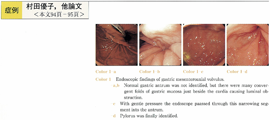
    A 66-year-old man was referred to our hospital because of recurrent attack of epigastralgia, epigastrial distension and nausea. Under the endoscopy, the gastric antrum was not identified at the first, then there were many convergent folds of gastric mucosa in the midstomach causing luminal obstruction. With gentle pressure the endoscope passed through this narrowing segment into the antrum. Upper GI series revealed upside down stomach and a diagnosis of gastric mesenteroaxial volvulus was made. Endoscopic correction of gastric volvulus was successfully performed using alpha-loop maneuver under fluoroscopy. The endoscope was inserted into the second portion of the duodenum to form an alpha-loop in the stomach. Then, the endoscope was torqued in a clockwise manner to allow uncoiling of the alpha-loop and correction of the gastric volvulus. This paper describes the classification, diagnosis and treatments of gastric volvulus and highlights the effectiveness of its endoscopic correction using an alpha-loop maneuver.
    Download PDF (601K)
  • Shizuka Nagaba, Masato Ooida, Satoshi Tanabe, Mitsuhiro Kida, Katsunor ...
    2001 Volume 58 Issue 2 Pages 96-97
    Published: June 15, 2001
    Released on J-STAGE: May 31, 2014
    JOURNAL FREE ACCESS
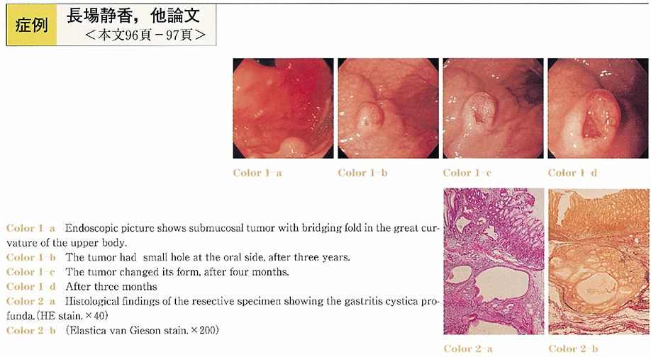
    Gastritis cystica profunda is rare, and it's natural course is still unknown. We report a 58-year-old man with gastraitis cystica profunda. We were able to follow up for four years. During the periods, its gross appearance and endoscopic ultrasonographic findings were changed in course of time. Initial endoscopic examination showed submucosal tumor. In addition to it, it revealed as solid tumor by ultrasonographic endoscopy. Three years later, cystic area appeared the tumor ultrasonographically. Partial gastric resection was performed because of severe gastrointestinal bleeding.
    It was diagnosed as gastritis cystica profunda by pathological examination finally.
    Download PDF (1293K)
  • Takayuki Ishii, Yoshihisa Namiki, Toru Furuya, Makoto Nakamura, Tateki ...
    2001 Volume 58 Issue 2 Pages 98-99
    Published: June 15, 2001
    Released on J-STAGE: May 31, 2014
    JOURNAL FREE ACCESS
    A 68-year-old man who had alcoholic liver cirrhosis with solitary gastric varices was admitted to our hospital. Gastric varices without gastro renal shunt was detected by arterio portography and 3 phase computed tomography. The patient was treated with an angiotensin II receptor antagonist, Losartan. As a results, reduced gastric varices was revealed by upper G-I endoscopy and decreased blood flow was observed by color doppler endoscopic ultrasonography. No symptomatic hyotensive reaction and no deterioration of liver or kidney function was observed. In this case an angiotensin II receptor antagonist was effective in treatment of solitary gastric varices.
    Download PDF (967K)
  • Noriko Tsuchiya, Takayoshi Nishino, Fumitake Toki, Hiroyasu Oyama, Syo ...
    2001 Volume 58 Issue 2 Pages 100-101
    Published: June 15, 2001
    Released on J-STAGE: May 31, 2014
    JOURNAL FREE ACCESS
    A 65-year-old female complaining of epigastric pain was admitted to our hospital. Laboratory data showed the elevation of biliary and pancreatic enzymes. The swelling and redness of papilla of Vater was observed by duodenoscope. The biopsy examination revealed adenocarcinoma. ERCP showed pancreas divisum, and the ventral pancreatic duct was dialated. EUS and IDUS showed a hypoechoic mass of about 20 mm diameter at the papilla of Vater, and the invasion into the pancreatic duct could not be excluded.
    Surgical material revealed papillary adenocarcinoma of 25mm in size showing no invasion into duodenum and pancreas. The surgical stage was stage I. We reviwed 6 cases of pancreas divisum concomitant with carcinoma of papilla of Vater in Japanese literature.
    Download PDF (777K)
  • Yuuich Takeda, Yumiko Hosoya, Tomoyuki Koyama, Keiichi Ono, Tsunehito ...
    2001 Volume 58 Issue 2 Pages 102-103
    Published: June 15, 2001
    Released on J-STAGE: May 31, 2014
    JOURNAL FREE ACCESS
    A-71-year-old man was admitted to our hospital because of abdominal pain and abdominal fullness due to duodenal stenosis. 6 months ago, he visited our hospital because of obstructive jaundice due to pancreas head carcinoma, and biliary stent was inserted following percutaneous transhepatic biliary drainage. On this admission, endoscopic and X-ray study showed severe stenosis at the 2nd portion of duodenum. An expandable metallic stent (EMS) was not enough long for passage through the narrowing space of duidenum. We make a new delivery system use of teflon tube. After cutting handlebar, teflon tube connected with outer tube of EMS. And the new delivery system was 150 cm length and detained at the senotic lesion. 5 days after treatment with EMS, he was able to take a diet for about 3 months. We think that EMS therapy for duodenal stenosis is useful to the improvement of quality of life.
    Download PDF (1524K)
  • Sonoko Suzuki, Kazuya Kawazoe, Tadashi Kitamura, Takashi Ohno, Teruo K ...
    2001 Volume 58 Issue 2 Pages 104-105
    Published: June 15, 2001
    Released on J-STAGE: May 31, 2014
    JOURNAL FREE ACCESS
    We reported a case of gastrointestinal stromal tumor (GIST) of duodenum in a young male.
    A 32-year-old male was admitted to our hospital because of right upper abdominal tumor. Abdominal CT revealed a tumor (10 cm in diameter) with necrosis developed extramurally from the second portion of the duodenum. Endoscopic examination revealed a tumor with easy bleeding ulcer of the second portion of the duodenum. Histopathological examination of biopsy specimens suggested that this tumor was a stroml tumor. Angiography revealed that this tumor was feeding with PSPD and ASPD.
    We performed pancreatoduodenectomy. Histopathologically, this tumor was composed of spindle cells arranged in interlacing-bundled pattern. Immunohistochemically, tumor cells were positive for CD34 and new myogenic markers (caldesmon and calponin) , but were negative for clasical myogenic markers (SMA, desmin) and Schwann cell markers (S-100 protein and Leu7) . Therefore, it was diagnosed as a duodenal stromal tumor with cajal cell and myogenic origin.
    We will need to study large number of patients to clarify diagnosis and malignant potential of GIST.
    Download PDF (675K)
  • Hideki Watanabe, Takaaki Ikeda, Tomoko Matumoto, Eriko Okada, Takeshi ...
    2001 Volume 58 Issue 2 Pages 106-107
    Published: June 15, 2001
    Released on J-STAGE: May 31, 2014
    JOURNAL FREE ACCESS
    A 60 years old woman was administered prednisolone (PSL) continuously because of autoimmune hepatitis (AIH) for 6 years. She was admitted to our hospital with purpuric rash on her extremities and hematochezia. We found erosion and multiple ulceration on terminal ileum by endoscopic examination of lower gastrointestinal tract and on the second portion of duodenum by endoscopic examination of upper gastrointestinal tract. We diagnosed anaphylactoid purpura from the biopsy of purpura and remarkable decline of factor XIII. She transiently got better without more administration (PSL was administrated as before) , but had recurrence of the disease. We administrated factor XIII concentrate to her for 3 days. She immediately got better after administration of factor XIII concentrate. In this case PSL may be resistant to the anaphylactoid purpura, because she was taken the disease during administration of PSL. And factor XIII concentrate may be effective to anaphylactoid purpura in adult.
    Download PDF (258K)
  • Miho Tabata, Yoshihiro Furuichi, Shuji Sugiki, Hiroko Mutsukawa, Masah ...
    2001 Volume 58 Issue 2 Pages 108-109
    Published: June 15, 2001
    Released on J-STAGE: May 31, 2014
    JOURNAL FREE ACCESS
    We experienced a case of a patient with Schönlein-Henoch purpura in whom small intestinal lesions were confirmed by colonoscopy.
    A 22-year-old male visited dermatology division, complaining purpura appeared in the bilateral lower legs. 8 days later be started bloody stool with abdominal pain and he was admitted. Colonoscopy revealed diffuse erosion with bleeding in the terminal ileum lesion. Cutaneous biopsy showed leukocytoclastic vasculitis was diagnosed Schönlein-Henoch purpura. Treatment was begun with steroids and DDS (diamino-diphenyl-sulphone) , which improved the symptoms.
    We investigated 68 reports of patients with Schönlein-Henoch purpura with apparent digestive lesions were confirmed during five years from 1996 to 2000. Although esophageal lesions were rarely found, erosions and ulcers were common findings in the second portion or lower region of the duodenum in many cases. Small intestinal ulcers are often diagnosed and in severe cases, operated cases are reported. Confirmation of small intestinal lesions by endoscopy was useful for the diagnosis of Schönlein-Henoch purpura complaining bloody stool.
    Download PDF (759K)
  • Hiroki Ochiai, Chi-Horng Shih, Keiichi Yoshino, Hiroaki Hattori, Shinj ...
    2001 Volume 58 Issue 2 Pages 110-111
    Published: June 15, 2001
    Released on J-STAGE: May 31, 2014
    JOURNAL FREE ACCESS
    A 49-year-old male was admitted to our hospital due to abdominal pain and nausea. Small bowel fluorography showed complete obstruction of the jejunum 30cm anal side from the ligament of Treitz. The endoscopic examination was performed with an intestinal fiberscope (Olympus X-SIF10) . There was an elevated lesion with granular and irregular surface similar to a villous tumor of the colon. The fiberscope did not pass the tumor to the anal side. Biopsy specimen showed moderately differentiated adenocarcinoma. Jejunal resection with lymph node dissection was performed followed by end to end anastomosis.
    There has been 50 cases of small intestinal cancer except duodenal cancer literally reported in Japan, which could be diagnosed preoperatively by intestinal endoscopy and our classification of those cancer is shown.
    Download PDF (715K)
  • Toshiro Sato, Hiroyuki Imaeda, Haruhiko Ogata, Yasushi Iwao, Yasuo Hos ...
    2001 Volume 58 Issue 2 Pages 112-113
    Published: June 15, 2001
    Released on J-STAGE: May 31, 2014
    JOURNAL FREE ACCESS
    A 30-year-old woman visited our hospital with complaints of abdominal pain, diarrhea and hematochezia in August, 1998. She often had raw fishes. Colonoscopic examination showed aphthas in the terminal ileum and she was diagnosed as aphthoid ileitis and treated by mesalazine. Her symptoms improved and colonoscopic examination showed improvement of aphthas in July, 1999. However, she again visited our hospital with complains of same symptoms in July, 2000. Colonoscopic examination showed a vivid anisakis larva penetrating the mucosa with redness and erosions in the terminal ileum. The symptoms disappeared after removal of it by colonoscopy with forceps. Relationship between aphthoid ileitis and intestinal anisakiasis is not clear. There has been only two cases of anisakiasis in the terminal ileum removed by colonoscopy including our case. It is important that anisakiasis-suspected case should undergo not only upper gastrointestinal endoscopy but also colonoscopy with precise observation of terminal ileum.
    Download PDF (310K)
  • Hiroyuki Oka, Shigeru Yamato, Masayuki Saruta, Shuuya Yoshinaga, Hirok ...
    2001 Volume 58 Issue 2 Pages 114-115
    Published: June 15, 2001
    Released on J-STAGE: May 31, 2014
    JOURNAL FREE ACCESS
    Retrograde ileography with colonoscope assisted insertion of balloon catheter into the terminal ileum is useful in diagnosing ileo-cecal and distal ileal lesions. However, this method takes much labor ; namely 1) intubation of colonoscope into distal ileum, 2) insertion of guide wire through the biopsy channel, 3) removal of colonoscope, 4) insertion of balloon catheter along guide wire, 5) injection of barium and air into the distal ileum. Moreover, the balloon catheter may slip out from distal ileum during studies. To avoid such labor and problems, we performed retrograde ileography without removing colonoscope and via a simple catheter which was inserted through biospy channel. This method is simple and quickly performed. Furthermore, it yields accurate information because the catheter positioning could be adjusted by manipulating colonoscope and the amount of barium and air in the distal ileum could be controlled freely.
    Download PDF (1245K)
  • Shigeo Yoshizawa, Tomoharu Yajima, Hiroshi Serizawa, Noriaki Watanabe, ...
    2001 Volume 58 Issue 2 Pages 116-117
    Published: June 15, 2001
    Released on J-STAGE: May 31, 2014
    JOURNAL FREE ACCESS
    A 27-year-old man was admitted to our hospital because of right lower abdominal pain. Abdominal CTscan showed a round mass 5cm in size consisted of fat tissue.
    Colonoscopic examination revealed a red-yellowish subpedunculated tumor about 5cm in diameter near Bauhin valve in the ascending colon.
    Colonoscopic partial resection was performed, and the tumor tissue revealed histologicaly benign lipoma. Five days after, therapeutic snaring for the remained tumor was done, and two months after, the tumor was disappeared with scarred mucosa. Two step endoscopic therapy, partial resection for histological diagnosis and subsequent snaring was safe and useful, for the giant lipoma.
    Download PDF (570K)
  • Kohei Kawakami, Mitsuhide Gotoh, Michio Hidaka, Takayuki Isikawa, Dais ...
    2001 Volume 58 Issue 2 Pages 118-119
    Published: June 15, 2001
    Released on J-STAGE: May 31, 2014
    JOURNAL FREE ACCESS
    54-year-old male had a two-day trip to Korea from March 24-26, 2000. He developed common cold-like symptoms from April 10, 2000. On April 15, the patient had a fever of 38-39℃, and his symptoms did not improve irrespective of medication. Diarrhea and bloody stool began on April 19, and he visited our hospital the following day. Colonoscopy performed immediately and revealed multiple undermining ulcers with peripheral mucosa showing an edematous elevation that located at the terminal ileum. And one of ulcer hemostais was carried out with henwclip device and local injective of HSE and clipping was conducted. After admission, the high fever continued. Blood cultures detected Salmonella typhi.
    Patients with high fever with bloody stool and colonoscopy reveals multiple undermining ulcers at the terminal ileum, it is important to suspect typhoid fever and to examine blood culture. There were 7 cases of typhoid fever conformed ideal ulcer by endoscope which is during there 30 years.
    Download PDF (206K)
  • Tateki Yamane, Hiroyuki Kato, Takayuki Ishii, Yoshihisa Namiki, Makoto ...
    2001 Volume 58 Issue 2 Pages 120-121
    Published: June 15, 2001
    Released on J-STAGE: May 31, 2014
    JOURNAL FREE ACCESS
    A 49-year-old man visited our hospital complaining diarrhea and lower abdominal pain. Abdominal CT scan examination showed thickened wall of the right sided colon, and colonoscopic examination revealed edematous and erosive mucosa and narrowed lumen in the intestine from the middle of the ascending colon to the terminal ileum. The biopsy specimen from the erosions showed non-caseous granulomas with Langhans giant cells, and it proved multiple bacillus bodies in the Ziehl-Neelsen stain. The feces specimen showed Gaffky-3 and the PCR method proved it as tubercle bacillus. Though chest roentgenogram showed no abnormality, chest CT scan examination revealed multiple granular shadows in the both lung. We thought it as miliary tuberculosis and suspected that intestinal tuberculosis was brought about hematogenously. It is rare that multiple tubercle bacillus are recognized in the biopsy specimen from the colonic lesion, so we reported it as this case.
    Download PDF (483K)
  • Itsuka Shosaku, Atsushi Kawaguchi, Masato Osaka, Kazumi Shimanouchi, N ...
    2001 Volume 58 Issue 2 Pages 122-123
    Published: June 15, 2001
    Released on J-STAGE: May 31, 2014
    JOURNAL FREE ACCESS
    A 21-year-old male had been suffering from ulcerative colitis (UC) from age 18. He was admitted to our hospital due to fever, abdominal pain, and frequent bloody stool. Although the general condition had been improved by intravenous steroid, frequent bloody stools developed and hypovolemic shock occurred at the 22nd hospital day. Urgent colonoscopy revealed local bleeding from an exposed vessel of the ulcer in the ascending colon, which was treated by endoscopic hemostasis by clipping. After the hemostasis, the dose of steroid could be tapered and general condition improved.
    In the case of UC with massive bleeding, urgent colonoscopy and endoscopic hemostasis shoud be attempted considering the possibility of local bleeding before urgent surgical therapy.
    Download PDF (241K)
  • Shin-ichi Takagaki, Kazuo Maruta, Naoki Shimizu, Morihito Igawa, Asako ...
    2001 Volume 58 Issue 2 Pages 124-125
    Published: June 15, 2001
    Released on J-STAGE: May 31, 2014
    JOURNAL FREE ACCESS
    A 43-year-old man had been suffering from recurrent attacks of bronchial asthma for 1 year. Two months prior to admission he reported aggravation of the symptoms, purpura on both his legs, weakened superficial sensation in his hands and fingers and watery stools, 8-times a day, accompanied by abdominal pain. He was admitted to our hospital to undergo careful examination. On admission, a peripheral blood test revealed high-level leukocytosis of 25,500/mm3 (eosinophiles, 57.3%) and increased IgE level of 2,990 U/mL. Based on these findings as well as the results of a skin biopsy, which showed eosinophilic vasculitis, a diagnosis of allergic granulomatous angitis (AGA) was made. Colon endoscopy revealed marginal flare with multiple erosions and ulceration in some parts of the sigmoid colon. After PSL therapy was initiated (60 mg/day) , the eosinophile count decreased to 5.1%, and the diarrhea and abdominal pain abated. Endoscopic examination performed after the treatment demonstrated a decreased number of mucosal erosions. PSL-dose tapering was stopped at 20 mg/day and, since no aggravation of symptoms was observed, the patient was discharged. In the present study, we report a case of colitis complicated with AGA we have encountered.
    Download PDF (703K)
  • Kentaro Sudo, Akira Hayasaka, Shin-ichi Ozawa, Tomoo Miyauchi, Takeshi ...
    2001 Volume 58 Issue 2 Pages 126-127
    Published: June 15, 2001
    Released on J-STAGE: May 31, 2014
    JOURNAL FREE ACCESS
    A 68-year-old man admitted to our hospital with the chief complaint of fever and right upper abdminal pain during the follow-up period of descending colon diverticulitis. Abdominal computed tomography and clinical laboratory findings suggested multiple liver abscess secondary to the colon diverticulitis. With our prompt antibiotics administration, the liver abscesses diminished and the patient's general condition was markedly improved. The part of the colon which had diverticulitis was resected to prevent the abscess recurrence.
    Liver abscess is one of the infrequent, but serious complications of diverticulitis. Early diagnosis and appropriate antibiotics treatment is believed to be a key to the better prognosis for such complicated patients. Both computed tomography and ultrasonography are powerful tools for the quick and non-invasive diagnosis.
    Download PDF (1103K)
  • Ken-ichi Komiya, Shin Tanaka, Kanako Higuchi, Syuka Mori, Masanori Yat ...
    2001 Volume 58 Issue 2 Pages 128-129
    Published: June 15, 2001
    Released on J-STAGE: May 31, 2014
    JOURNAL FREE ACCESS
    A 61-year-old man visited our hospital for melena. Colonoscopy revealed colonic diverticulosis and massive bleeding in the ascending colon, but the location of bleeding diverticula was not determined. Mesenteric angiography was then carried out. A superior mesenteric arteriogram failed to demonstrate the bleeding site, but selective right colic arteriogram showed extravasation of contrast medium in the ascending colon. The patient was successfully treated with subsequent transcatheter arterial embolization. In the present case, initial colonoscopy was useful in evaluating bleeding site, which enabled successful selective angiography and arterial embolization.
    Download PDF (1213K)
  • Tsuneo Ishitsuka, Kazutomo Togashi, Masaki Okada, Fumio Konishi, Hideo ...
    2001 Volume 58 Issue 2 Pages 130-131
    Published: June 15, 2001
    Released on J-STAGE: May 31, 2014
    JOURNAL FREE ACCESS
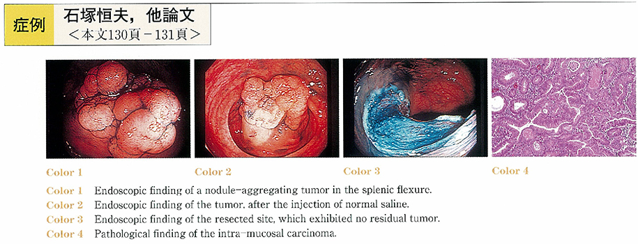
    The case was a 53-year-old male who complained of a fecal occult blood. Colonoscopy revealed a nodule-aggregating tumor of 49 mm in diameter in the splenic flexure. In supine position, it was not possible to have the entire view of the lesion. However, in left lateral position, it was possible to have an entire view of the lesion. We injected normal saline containing epinephrine into the submucosa adjacent to the oral side of the tumor. That procedure resulted in a protrusion of the oral side of the tumor. Subsequently, the anal side of the tumor was pressed down with the base of the snare, which enabled a successful snaring of the lesion. After first resection, a small part of the tumor remained at the oral side. The residual tumor was resected completely by the second snaring. Histologically, the tumor was a tubulo-villous adenoma containing the areas of well-differentiated adenocarcinoma not invading to the submucosal layer. Colonoscopy was repeated three months after the resection, which revealed no residual or recurrent tumors.
    It has been reported that the maximum size of flat colorectal tumor, in which en bloc endoscopic mucosal resection can be performed, is 30mm in diameter. We present a case of a large flat tumor totally resected endoscopically almost in one piece.
    Download PDF (622K)
  • Hidejiro Kawahara, Makoto Ohno, Hirotosi Ishikawa, Katsuya Hirai, Teru ...
    2001 Volume 58 Issue 2 Pages 132-133
    Published: June 15, 2001
    Released on J-STAGE: May 31, 2014
    JOURNAL FREE ACCESS
    A 54-year-old male visited our hospital as an emergency patient due to severe epigastric pain. About 20 years ago he received distal partial gastrectomy due to duodenal ulcer. Endoscopic cholangiography with straight view scope demonstrated that he had several CBD stones of 10 or more mm in diameter. So he underwent a surgical operation. But a residual CBD stone was found by post operative cholangiography from C tube. The residual stone was successfully removed by EST with straight view scope.
    It is very difficult with side view scope to approach the afferent loop in the case of partial gastrectomy with Billroth 2 reconstraction. However, a straight view scope may be the tool of the first line for trying EST in such a case.
    Download PDF (1449K)
  • Katsushi Seza, Fumitaka Nagashima, Tsunehiro Endo
    2001 Volume 58 Issue 2 Pages 134-135
    Published: June 15, 2001
    Released on J-STAGE: May 31, 2014
    JOURNAL FREE ACCESS
    The case described a 75-year-old man who has been treated because of liver cirrhosis due to hepatitis C. In May 2000, a common bile duct (CBD) stone, measuring 8 mm in diameter, was detected at abdominal CT scanning. Because he had undergone Billroth II gastrectomy, it was difficult to access to a papilla and insert a catheter into the bile duct using a side-viewing scope or a direct-viewing scope. An anterior oblique-viewing make access of Vater's papilla and control of a catheter easy, but the cannulation was failed. Thus percutaneous transhepatic cholangiodrainage was performed and an anterior oblique-viewing endoscope was inserted. After that, a guidewire was introduced from drainage tube into the scope ; The wire was grasped by a forceps and was passed through the forceps channel ; endoscopic papillary balloon dilation (EPBD) was then performed using a balloon catheter for dilation (MaxForce ; maximum dilated diameter 8 mm ; length 3 cm) , leading to successful removal of stones. Our experience in this case indicated that EPBD with an anterior oblique-viewing endoscope is useful for the treatment of CBD stones following Billroth II gastrectomy, and that EPBD can be applied with the percutaneous transhepatic assistance, even to the cases with difficulty in transpapillary approach, demonstrating the potent application of EPBD to a wider range of cases.
    Download PDF (1048K)
  • Satoru Gotoh, Masahiko Murakami, Kouji Otsuka, Akira Ishihara, Yoshihi ...
    2001 Volume 58 Issue 2 Pages 136-137
    Published: June 15, 2001
    Released on J-STAGE: May 31, 2014
    JOURNAL FREE ACCESS
    Torsion of the gallbladder is a relatively rare disease. It has been reported to be hard to diagnose preoperatively. We report a case of torsion of the gallbladder diagnosed preoperatively with striking finding on MRCP.
    An 85 years old female admitted to our hospital because of severe abdominal pain and vomiting. She was diagnosed acute cholecystitis and chololithiasis by findings on computed tomography and ultrasonogram. PTGBD and MRCP were performed. MRCP showed a tapering (smooth) interruption both side of the cystic duct. Because of this striking finding on MRCP, she was diagnosed torsion of the gallbladder preoperatively and underwent laparoscopic cholecystectomy. In the operation, the gallbladder was twisted clock wisely about 180 degrees, and noted as a Gross type II, floating gallbladder.
    In this case, MRCP revealed a tapering (smooth) interruption both side of the cystic duct. We consider that this striking finding is useful for diagnosis to distinguish torsion of the gallbladder from gallbladder (choledochal) stone and gallbladder cancer.
    Download PDF (934K)
feedback
Top